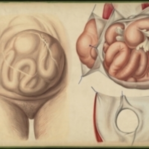
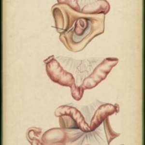
After Jean Cruveilhier's Anatomie pathologique du corps humain, vol. 2, liv. 24, pl. 6 Large watercolor showing several views of umbilical hernias. The leftmost is an external view showing the lower abdomen and groin of a woman suffering from an extreme umbilical hernia. The form of the intestines can be seen bulging through the skin, and white stretch marks cross the abdomen. The upper right is an interior view of an umbilical hernia, showing the pink intestines surrounded by white abdominal tissue, which is pulled away with blue hooks. The lower right image is of the large circular hole in through which the hernia protruded. Watercolor is framed in green sewn textile, with metal grommets in each of the four corners.
The Harvard Medical Library does not hold copyright on all the materials in the collection. For use information, contact the Warren Anatomical Museum Curator at chm@hms.harvard.edu
Contact host institution for more information.
Notes:
Henry Jacob Bigelow employed artist Oscar Wallis exclusively from 1848 - 1854 to paint a series of large teaching watercolors to illustrate Bigelow's lectures at Harvard Medical School. Wallis painted the teaching diagrams from local subjects and from the atlases of established medical authorities. The effort cost Bigelow $6,000. In 1890 Bigelow presented the watercolors to Reginald H. Fitz to be used in the Harvard Medical School's Department of Anatomy. The watercolors were transferred into the Warren Anatomical Museum between 1890 and 1930.