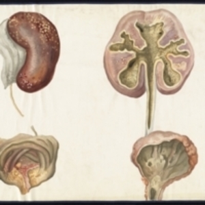
Teaching watercolor of interior and exterior of diseased kidney and bladder
Description:
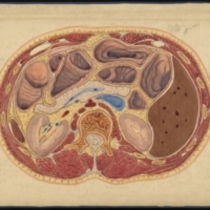
Large watercolor of diseased kidney and bladder. On left, the exterior of the red-brown kidney has yellow and pink dots, and bladder below is painted in green and pink, with yellow dots. On right, the interior of the kidney is painted with olive green, and yellow dots lined in red are scattered throughout the pink of the kidney organ, and the inside of the bladder is gray. Watercolor is framed in blue sewn textile.
The Harvard Medical Library does not hold copyright on all the materials in the collection. For use information, contact the Warren Anatomical Museum Curator at chm@hms.harvard.edu
Contact host institution for more information.
Notes:
The watercolor was painted by William J. Kaula in 1896 for John Collins Warren to use in teaching. The watercolor was left in the Harvard Medical School Department of Anatomy after Warren's death in 1927. The Department donated the watercolors to the Warren Anatomical Museum on 05/02/1929.