Teaching watercolor of rectal disease and neoplasms
Description:
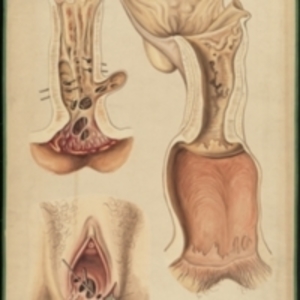
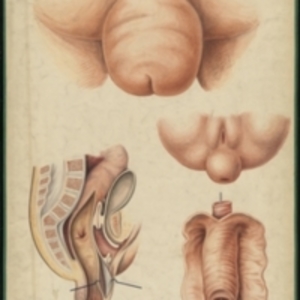
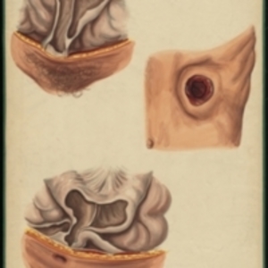
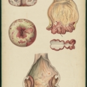
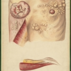
After Jean Cruveilhier's Anatomie pathologique du corps humain, vol. 2, liv. 33, plate 1 Large watercolor depicting diseases of the rectum. On the left are two cases of rectal inflammation that simulate cancer of the rectum. Views are from a cross section of the rectum, and from the exterior of a female patient. Metal pins are inserted to show the perforations of the tissue. On the right is rectal cancer, with tissues drawn in tan and pink. Watercolor framed in green sewn textile, with metal grommets in each of the four corners.
The Harvard Medical Library does not hold copyright on all the materials in the collection. For use information, contact the Warren Anatomical Museum Curator at chm@hms.harvard.edu
Contact host institution for more information.
Notes:
Henry Jacob Bigelow employed artist Oscar Wallis exclusively from 1848 - 1854 to paint a series of large teaching watercolors to illustrate Bigelow's lectures at Harvard Medical School. Wallis painted the teaching diagrams from local subjects and from the atlases of established medical authorities. The effort cost Bigelow $6,000. In 1890 Bigelow presented the watercolors to Reginald H. Fitz to be used in the Harvard Medical School's Department of Anatomy. The watercolors were transferred into the Warren Anatomical Museum between 1890 and 1930.